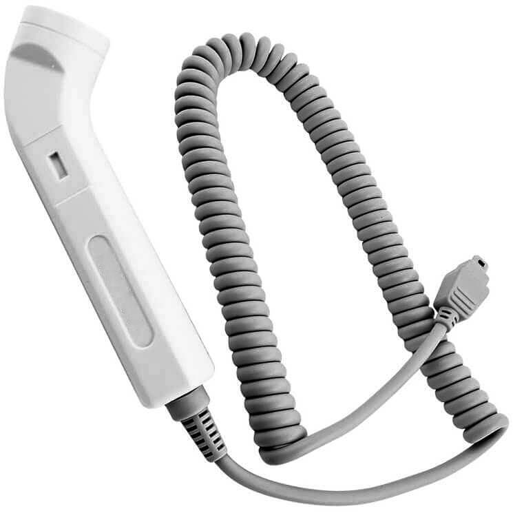
Doppler Cord For Fetal Doppler And Vascular Doppler Probe From 2Mhz To 8Mhz
In medical cables,especially coil cables,AED cable and doppler cord are the two main forms custom doppler cord for doppler probe, 2Mhz to 8Mhz is the main frequency in Fetal Doppler And Vascular Doppler Probe . higher Frenquency for more peripheral the detection will be.Choose the right doppler cord for your doppler probe,you will get more accurate detection.With the correct matchable probes, Many dopplers have the ability to assess fetal heartbeats and what gestation periods or will you be assessing vascular blood flow.(in this sense,it is a fetal doppler and vascular doppler at the same time)
How to Choose a right probe for doppler ?
Doppler cord is not equal to doppler probe,ususally a doppler probe compose a detector and a suitable cord whose frequency is suitable for the detector. As the dectors are usually 3-8 MHZ (≥3MHZ is called High Frequency)),it is meanningless to mention High Frequency here. Vascular and obstetrical probes work on different ultrasound formats and it is highly improbable that you will be able to effectively assess the opposite sounds with accuracy to which the probe is designed.
Obstetrical applications.
With hand held dopplers, all probes less than a 3MHz will be used for obstetrical application.
2MHz fetal doppler probe– This is a later term obstetrical probe and will attain deeper penetration. This is often the choice for anything after 21 weeks gestation. It is also often available in a waterproof version for tub labor or assessments in the shower.
3MHz fetal doppler probe– This is the early term obstetrical probe. It is more peripheral than the 2MHz and often used in the office assessments. Some have found that this probe is well suited during delivery once the child has dropped into position for birthing.
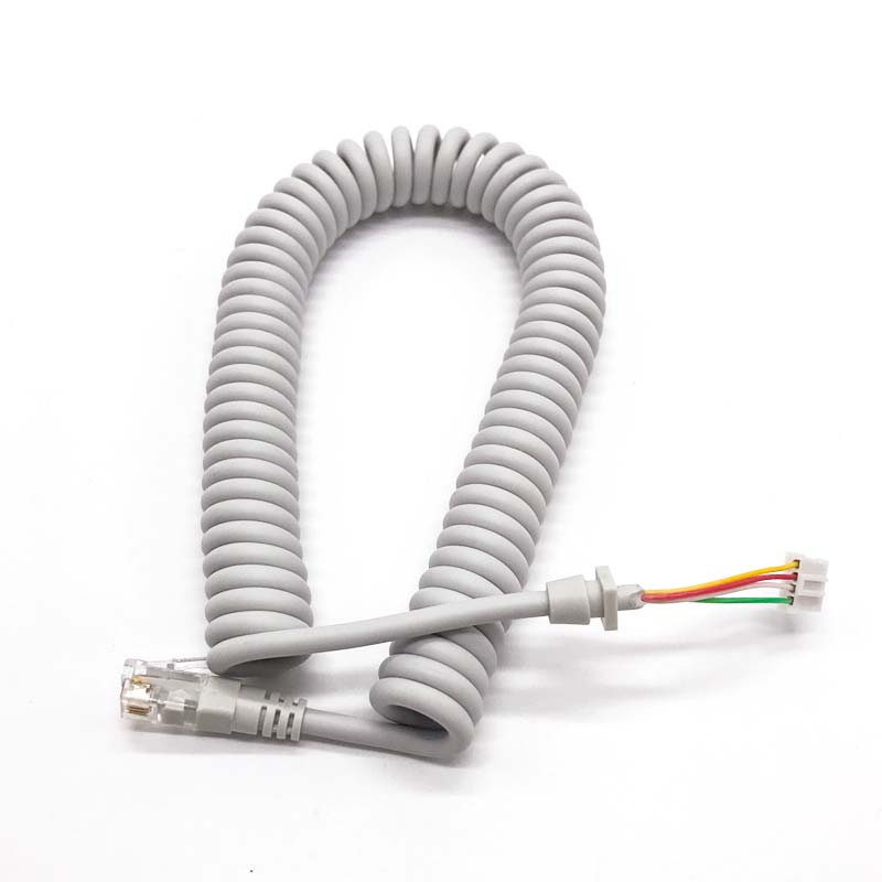
Raw Doppler cord
custom coil doppler cord for different frequency purpose
3mhz Doppler probe cable
Vascular applications
Everything above a 4MHz will be used for vascular applications.
- 4MHz – This is a very deep penetration vascular probe that is best suited to areas requiring assessment of deep arterial sounds such as aortic, femoral and other major vascular assessments.
- 5MHz – This probe is commonly paired with the 8MHz probe as a good selection for total vascular assessments. This probe will be great for assessing with bariatrics or in some cases, the sclerosis patients. It is a mid-level assessment and a good multi-purpose probe to have.
- 8MHz – This is the most popular probe for peripheral detection such and ABI (Ankle, Brachial Index) assessments. Those looking for pedal pulses, brachial pulses and digital pulses will prefer the 8MHz. Some Dopplers will offer this in a Sterilizable model. These are often used intra-operatively.
Doppler probe Clinical application based on different frequency range
- Small lesions of chest wall, pleura and lung periphery: 7-7.5MHz linear array probe or convex array probe
- Liver ultrasound examination:
①Convex array probe or linear array probe
②Adults: 3.5-5.0MHz, children or thin adults: 5.0-8.0MHz, obese people: 2.5MHz
- Gastrointestinal ultrasound examination:
①The convex array probe is used for abdominal examination, the frequency is 3.5-10.OMHz, generally 3.5-5.0MHz is the most commonly used
②Intraoperative ultrasound: 5.0-12.0MHz parallel linear array probe
③Endoscopic ultrasound: 7.5-20MHz
④Intrarectal ultrasound: 5.0-10.0MHz
⑤Ultrasound guided puncture probe: 3.5-4.OMHz, micro convex probe, small phased array probe with puncture guide frame.
5. Kidney ultrasound: phased array, convex array or linear array probe, 2.5-7.0MHz; children can choose higher frequency 6.Ultrasound examination of retroperitoneum: convex array probe: 3.5-5.OMHz, for those who are small and thin, the high-frequency probe of 7.0-10.0 can be used.
7.Adrenal ultrasound: convex array probe is preferred, 3.5MHz or 5.0-8.OMHz
8.Brain ultrasound: two-dimensional 2.0-3.5MHz, color Doppler 2.0MHz
9.Jugular artery and vein: linear array or convex array probe, 5.0-10.OMHz
10.Vertebral artery: 5.0MHz
11. Bone joint soft tissue ultrasound: 3.5MHz, 5.0MHz, 7.5MHz, 10.OMHz
12 Extremity vascular ultrasound: linear array probe, 5.0-7.5MHz
13.Eyes: ≥7.5MHz, preferably 10-15MHZ
14.Ultrasound of parotid gland, thyroid gland and testis: 7.5-10MHz, linear array probe
15.Breast ultrasound: 7.5-10MHz, without high-frequency probe, 3.5-5.OMHz probe can be used to add water bladder 16.Parathyroid ultrasound: linear array probe, above 7.5MHz